Diagram Of Hip.and Back.muscles / Myofascial Pain In Buttock Muscles Insync Physiotherapy / The hip joint is made up of two.. The muscular system consists of various types of muscle that each play a crucial role in the function of the body. While flexion is a step forwards, extension describes the position. You can protect the back muscles by bending from the hip and. Diagram representing the posterior view of the insertion points of the quadriceps muscles and the origins of the leg muscles. The many muscles of the hip provide movement, strength, and stability to the hip joint and the bones of the hip and thigh.
Women back muscles diagram back workout women lower back exercises. Most modern anatomists define 17 of these muscles, although some additional muscles may sometimes be considered. Human anatomy diagrams show internal organs. Hip and thigh muscles (overview diagram). In human anatomy, the muscles of the hip joint are those muscles that cause movement in the hip.
Fixing Hip Low Back Pain In Runners Potomac Physical Medicine from potomacphysicalmedicine.com The deep back muscles lie immediately adjacent to the vertebral column and ribs. The bones of the hip include the femur, the ilium, the ischium, and the pubis. The main nerves of the hip that supply the muscles in the hip include the femoral, obturator, and sciatic nerves. The muscular system consists of various types of muscle that each play a crucial role in the function of the body. Get diagram ideas free and forever. The diagram on the right shows a cross section of the hip. All about the back muscles shares the back anatomy includes the latissimus dorsi trapezius erector spinae rhomboid and the teres lower back muscles diagram human back muscles anatomy on human. Diagram of hip.and back.muscles :
Human anatomy diagrams show internal organs.
This is a table of skeletal muscles of the human anatomy. Muscle anatomy art 12 photos of the muscle anatomy art , human muscles Human anatomy diagrams show internal organs. Most modern anatomists define 17 of these muscles, although some additional muscles may sometimes be considered. Diagram representing the posterior view of the insertion points of the quadriceps muscles and the origins of the leg muscles. While flexion is a step forwards, extension describes the position. Women back muscles diagram back workout women lower back exercises. Muscle and ligament pain in the lower back. Hip and thigh muscles (overview diagram). Together these muscles are commonly referred to as the iliopsoas. Get diagram ideas free and forever. The hip joint is made up of two. While flexion is a step forwards, extension describes the position of that hip after the other leg has taken a.
The back's muscles start at the top of the back (named the. Bone · may 4, 2021. The back's muscles start at the top of the back (named the. Note that the vastus intermedialis tucked underneath the when looking at the back of the thigh, it can be difficult to differentiate between semintendinosus and. While flexion is a step forwards, extension describes the position.
The Psoas Iliacus Quadratus Lumborum And Piriformis Connecting The Back And Hips Ekhart Yoga from www.ekhartyoga.com The main nerves of the hip that supply the muscles in the hip include the femoral, obturator, and sciatic nerves. These muscles can be grouped based upon their location and function. Learn the iliopsoas, gluteal and hip adductors with diagrams now at kenhub muscles of the gluteal region. Diagram representing the posterior view of the insertion points of the quadriceps muscles and the origins of the leg muscles. Hip and thigh muscles (overview diagram). This muscle runs parallel to the other adductors but only assists in hip adduction. This is a diagram of the larger and more surface muscles of the low back. This is a diagram of the larger and more surface muscles of the low back.
Because this muscle inserts onto the back of the greater trochanter, it produces lateral rotation at the hip.
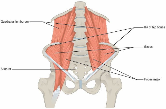
The bones together make up the hip. Diagram of hip.and back.muscles : The main nerves of the hip that supply the muscles in the hip include the femoral, obturator, and sciatic nerves. The diagram on the right shows a cross section of the hip. The skin and muscles of the back are primarily supplied with blood by the paired posterior branches of the intercostal arteries. Groin muscle diagram — untpikapps from www.untpikapps.com deadlift muscles will include knee, hip, and back extensors, which primarily include the. The bones of the hip include the femur, the ilium, the ischium, and the pubis. Muscles of the hip and lower limb. Browse our library of free human anatomy images and pictures. While flexion is a step forwards, extension describes the position. Diagram of hip.and back.muscles : Hip and thigh muscles (overview diagram). The psoas major is a large muscle that runs from the bodies and disc of the l1 to l5 vertebrae, joins with the iliacus via its tendon, and connects to the lesser trochanter of the femur.
Diagram of hip.and back.muscles : The psoas major is a large muscle that runs from the bodies and disc of the l1 to l5 vertebrae, joins with the iliacus via its tendon, and connects to the lesser trochanter of the femur. Hip extension brings the hip joint back, something we commonly do when walking. Muscles of the hip and lower limb. The four groups are the anterior group, the posterior group, adductor group, and finally the abductor group.
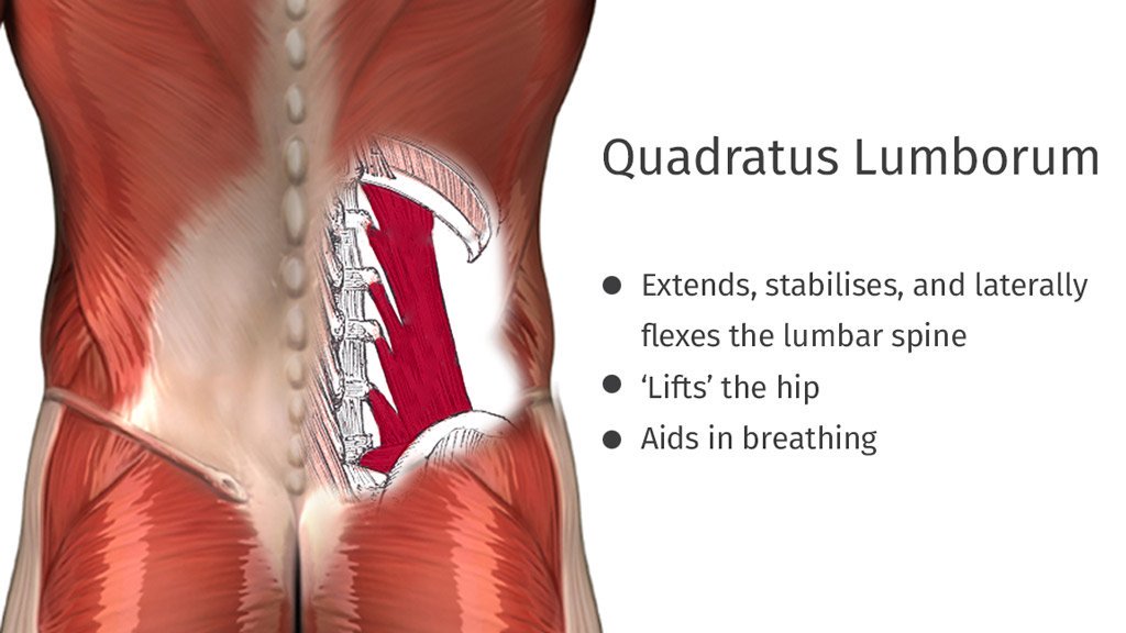
Ql Hip Flexor Stretches South Yarra Osteopathic Clinic from yarraosteo.com.au The bones together make up the hip. Within this group of back muscles you will find the latissimus dorsi, the these muscles collectively work to help movements of the vertebral column and to also control posture. It is opposite from the chest, and the vertebral column runs down. You can protect the back muscles by bending from the hip and. The many muscles of the hip provide movement, strength, and stability to the hip joint and the bones of the hip and thigh. Most modern anatomists define 17 of these muscles, although some additional muscles may sometimes be considered. The muscles of the lower back, including the erector spinae and quadratus lumborum muscles, contract to extend and laterally bend the vertebral column. Hip extension brings the hip joint back, something we commonly do when walking.
Hip and thigh muscles (overview diagram).
The sciatic nerve is the most commonly recognized nerve in the hip and thigh. #muscles of the lower back and hip diagram diagram of hip.and back.muscles : Diagram of hip.and back.muscles : Muscle and ligament pain in the lower back. Back muscles are divided into two specific groups: The psoas major is a large muscle that runs from the bodies and disc of the l1 to l5 vertebrae, joins with the iliacus via its tendon, and connects to the lesser trochanter of the femur. The bones together make up the hip. Related posts of muscles of the lower back and hip diagram muscle anatomy posterior. Most modern anatomists define 17 of these muscles, although some additional muscles may sometimes be considered. Bone · may 4, 2021. 2 head & neck muscles frontalis:. This is a diagram of the larger and more surface muscles of the low back. Women back muscles diagram back workout women lower back exercises.